Gamekeeper’s Thumb
What is Gamekeeper’s Thumb?
Ligaments are tough bands of fibrous tissue that connect bones together to form a joint. Collateral ligaments are important structures found on either side of each finger and thumb joint that prevent abnormal side-to-side bending of the joint. The main knuckle joints are formed by the connections of the finger bones (phalanges) to the hand bones (metacarpals). These joints found at the base of the fingers are called the metacarpophalangeal joints (MCP joints). The MCP joints work like a hinge when you bend and straighten your fingers and thumb.
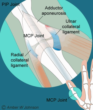
In the thumb, the collateral ligaments attach to the middle of the metacarpophalangeal joint and help to stabilize the thumb. The ulnar collateral ligament (UCL) is located on the inside of the thumb near to the web space of the hand, while the radial collateral ligament is located on the outside of the thumb (Figure 1).

Figure 1
Injury to the ulnar collateral ligament is fairly common. Acute UCL injuries most often occur from a fall on an outstretched hand with the thumb extended. This injury is frequent in skiers who fall while gripping a planted ski pole, and has thus been coined “Skier’s thumb”. Injury to the UCL is also called “Gamekeeper’s thumb” as it was an injury common to Scottish gamekeepers from repetitive forces to the thumb MCP joint while harvesting rabbits. Today, the terms “Skier’s thumb” and “Gamekeeper’s thumb” both are indicative of injury to the UCL.
Signs and Symptoms:
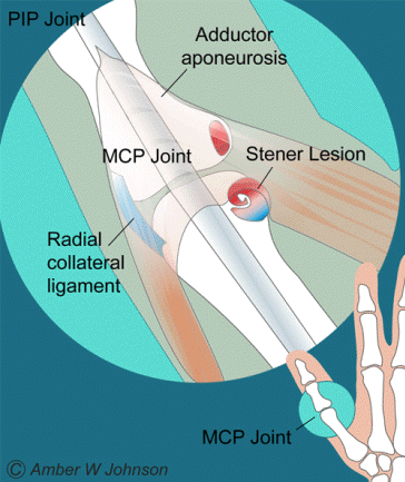
Acute injury to the ulnar collateral ligament is characterized by painful swelling and weakness when grasping with the thumb. Bruise-like discoloration of the skin may be present around the thumb. In a complete tear, the loose end of the torn UCL may retract and form a bump that can be felt along the edge of the thumb near the palm of the hand. This bump, called a “Stener lesion,” makes it difficult to hold or pinch things between your thumb and index finger. A Stener lesion forms when the ruptured end of a torn UCL becomes entrapped by the fascial extension of the adductor muscle (called the adductor aponeurosis), which inhibits the UCL from healing on its own (Figure 2).

Figure 2
Conservative Options:
Non-surgical treatment can be considered for a partially torn UCL if a Stener lesion is not present, and if the thumb joint is fairly stable. This indication will usually heal without surgery if treated early. Conservative treatment will consist of an initial period of rest with a cast or brace (usually a 4 week immobilization period), along with taking non-steroidal anti-inflammatory medications and applying ice to the thumb daily until the pain and swelling are gone. Immobilization is followed by an additional 2-week period of splint protection, during which active range of motion exercises are initiated. Healing with full recovery may be expected with a partial tear because the injured ligament maintains its normal anatomic relationship. However, strenuous activity with the thumb should still be avoided for 3 months following the injury. Patients commonly experience a degree of aching pain on the palm side of the thumb joint for 6 or more months after the injury even though the thumb joint is stable upon examination. Athletes with minor injuries to the UCL may expect to return to play wearing a protective ‘playing splint’ after the thumb has been completely immobilized for 2-4 weeks. The goals of nonsurgical treatment of a torn UCL are to provide healing and pain relief to the patient, and to restore stability to the MCP joint.
The presence of a Stener lesion may be difficult to diagnose without the use of MRI or ultrasound technology. However, a Stener lesion is imperative to diagnose, as a missed Stener lesion that is non-operatively treated predictably leads to prolonged dysfunction and pain. If a Stener lesion is present, surgical repair is usually necessary to reduce pain and to stabilize the thumb.
Surgical Options:
Surgical treatment is indicated when a partial tear fails to respond to conservative treatment, when there is a complete rupture, when there is significant instability, or when a piece of bone fragments off the main bone (an avulsion fracture). In general, the best functional results are obtained when surgical correction is achieved within 3 weeks of the initial injury.
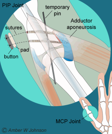
The surgeon will make a small incision over the back of the MCP joint of the thumb and will expose the MCP joint, the adductor aponeurosis, and Stener lesion (if present). The repair is made by releasing the Stener lesion from the adductor aponeurosis, extending the UCL to its original length, then anchoring the ligament back to the bone with stitches. The repair will be temporarily reinforced by placement of a single pin across the joint, and with sutures passed through the proximal phalanx attached to a button fixture on the external side of the thumb (Figure 3). This fixture will remain in place until adequate healing has occurred (about 4 weeks), returning stability to the MCP joint.

Figure 3
Dr. McNamara’s Post Op Protocol
Dr. Gray’s Post Op Protocol
Note: These instructions are to serve as guidelines and are subject to Physician discretion. Actual progress may be faster or slower depending on the individual.
